
 Fax: (239) 692-9436
 Tel: 239-430-3668
Adult Acquired Flatfoot (PTTD): Why the Arch Collapses, and How We Rebuild It
Posterior tibial tendon dysfunction (PTTD) is the most common cause of adult acquired flatfoot deformity — a slow, progressive collapse that can end in a rigid, arthritic hindfoot if it isn’t caught early. Dr. Kevin Lam reconstructs advanced cases with a minimally invasive triple arthrodesis technique built for a faster, less painful recovery.
What Is Posterior Tibial Tendon Dysfunction?
The posterior tibial tendon is the primary sling holding up the arch of the foot. When it degenerates from chronic overload, the tendon stretches and loses mechanical advantage. The arch settles, the heel rolls into valgus, and the forefoot abducts out from under the leg. Left untreated, a flexible, correctable flatfoot can progress into a fixed, arthritic deformity that no longer responds to bracing or physical therapy — this progression is what surgeons refer to as adult acquired flatfoot deformity (AAFD).
Who Gets PTTD — and Why Weight Matters
Far More Common in Women
Symptomatic PTTD is reported at a markedly higher rate in women than men, typically presenting in the fifth through seventh decade of life.
Obesity Is an Independent Risk Factor
Published case-control studies repeatedly identify elevated body mass index as an independent risk factor for PTTD — distinct from age, sex, hypertension, and diabetes, which also raise risk.
The Mechanical Link
Every step loads the posterior tibial tendon with a multiple of body weight. Excess weight raises both the baseline strain on the tendon and the ground-reactive force driving arch collapse with every stride.
Staging PTTD: The Johnson & Strohm Classification
Every flatfoot reconstruction starts with staging under the classification originally described by Johnson and Strohm, later extended with a fourth stage to account for deltoid ligament involvement. Staging determines whether a foot can still be treated with tendon repair and soft-tissue correction, or whether it has progressed to a fixed deformity that requires joint fusion.
STAGE I
Tenosynovitis — Flexible, Tendon Intact
Pain and swelling with no visible deformity. The tendon is inflamed but structurally intact and normal in length. The arch is still present, and the patient can typically perform a single-limb heel rise, though it may be painful.
STAGE II
Tendon Elongation — Flexible Flatfoot Deformity
The tendon is elongated and degenerated. Forefoot abduction produces the classic “too-many-toes” sign from behind, and the patient can no longer perform a single-limb heel rise. The deformity is still passively correctable, so reconstruction is usually soft-tissue based — tendon transfer with or without an osteotomy.
STAGE III
Fixed Deformity — Rigid, Arthritic
Chronic malalignment leads to secondary arthritic change in the subtalar and talonavicular joints. The deformity is now fixed rather than flexible. This is the stage at which joint-sparing procedures give way to arthrodesis (fusion) as the reliable option.
STAGE IV
Ankle Involvement — Later Addition
Added to the original three-stage system to describe cases where chronic hindfoot valgus stretches the deltoid ligament, tilting the talus within the ankle mortise. Reconstruction planning must then account for the ankle joint in addition to the hindfoot.
Case Study: Reading the Collapse on X-Ray
These weight-bearing films are from an actual FFLC Stage III–IV reconstruction in an overweight female patient with longstanding PTTD.
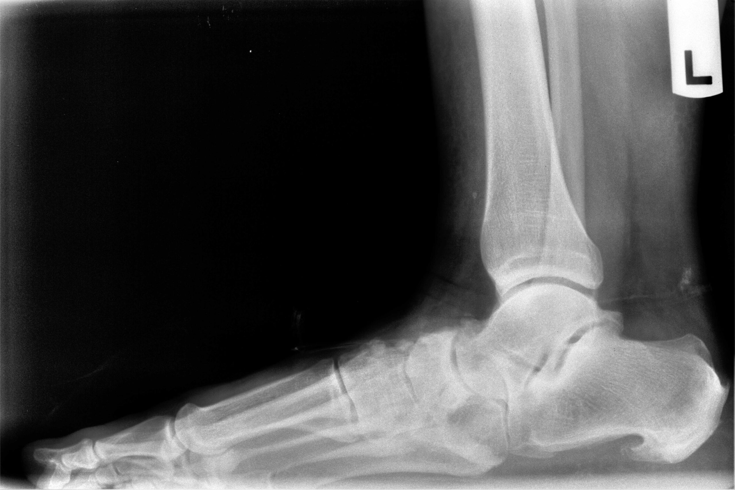
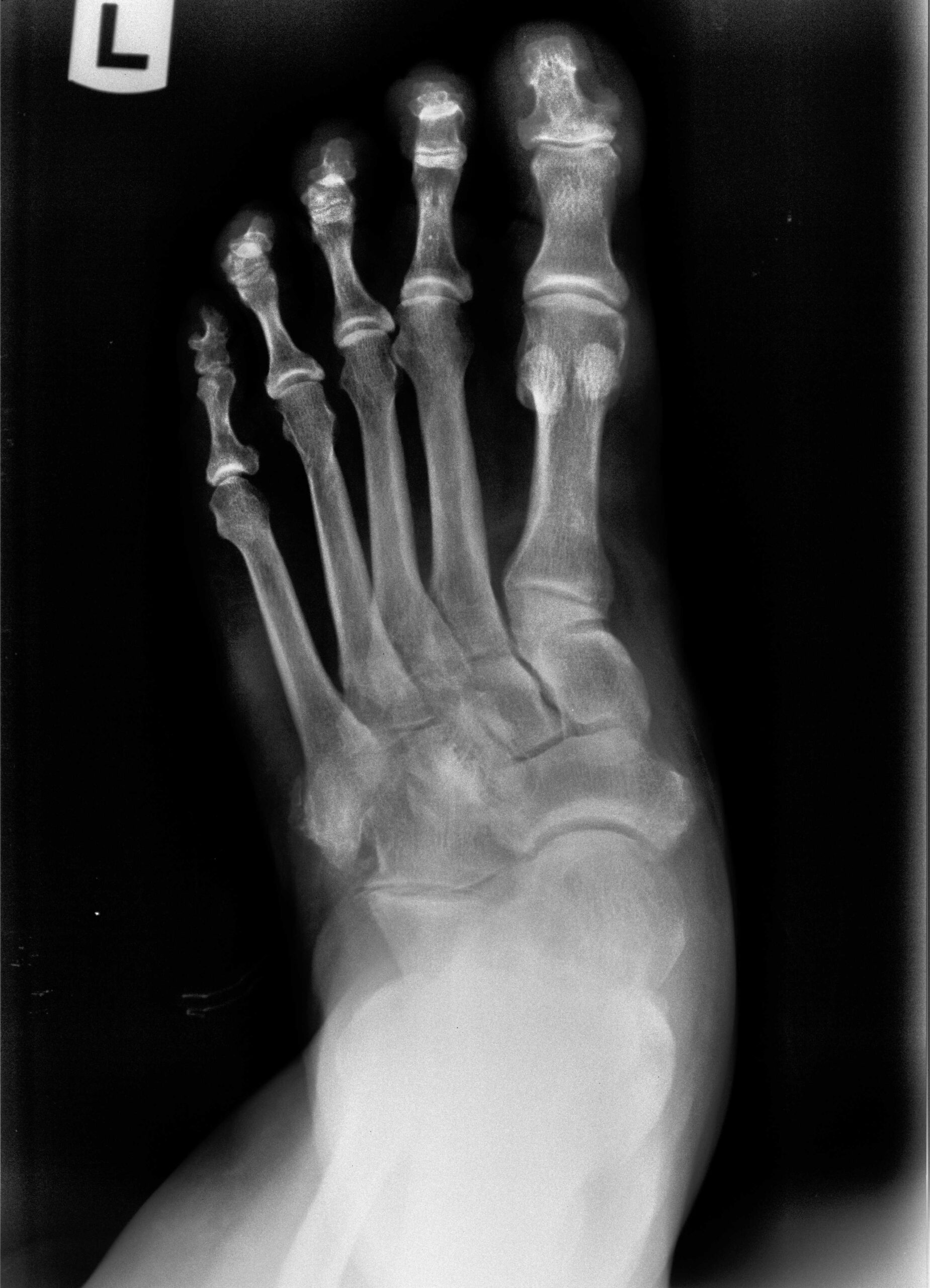
Lateral view: the talus has dropped into plantarflexion, increasing the talar declination angle, with flattening of the calcaneal pitch. AP view: the navicular has drifted laterally off the talar head, with loss of talar head coverage and forefoot abduction. Together these findings are consistent with a fixed, arthritic Stage III–IV adult acquired flatfoot deformity, no longer amenable to soft-tissue reconstruction alone.
The Procedure: Triple Arthrodesis with Minimally Invasive Subtalar Dowel Fusion
For fixed, arthritic Stage III–IV deformities, Dr. Lam’s preferred approach is a triple arthrodesis — fusing the subtalar, talonavicular, and calcaneocuboid joints to permanently realign and stabilize the hindfoot. Where joint anatomy allows, he performs the subtalar fusion using a dowel technique: a cylindrical reamer prepares and fuses the joint through a small sinus tarsi portal rather than a full open arthrotomy, meaning less soft-tissue stripping, less devascularization, and a smaller wound — the basis for a faster early recovery compared with a fully open triple arthrodesis.
01 — Realign
The talonavicular joint is reduced to re-cover the talar head and correct forefoot abduction, restoring structural alignment before any joint is fixed in place.
02 — Fuse
A dowel-shaped reamer prepares a cylindrical fusion surface at the subtalar joint through a limited portal, instead of opening the entire joint.
03 — Fix
Screw fixation compresses the subtalar joint while staples secure the talonavicular joint, holding the corrected position rigid while the fusion consolidates.
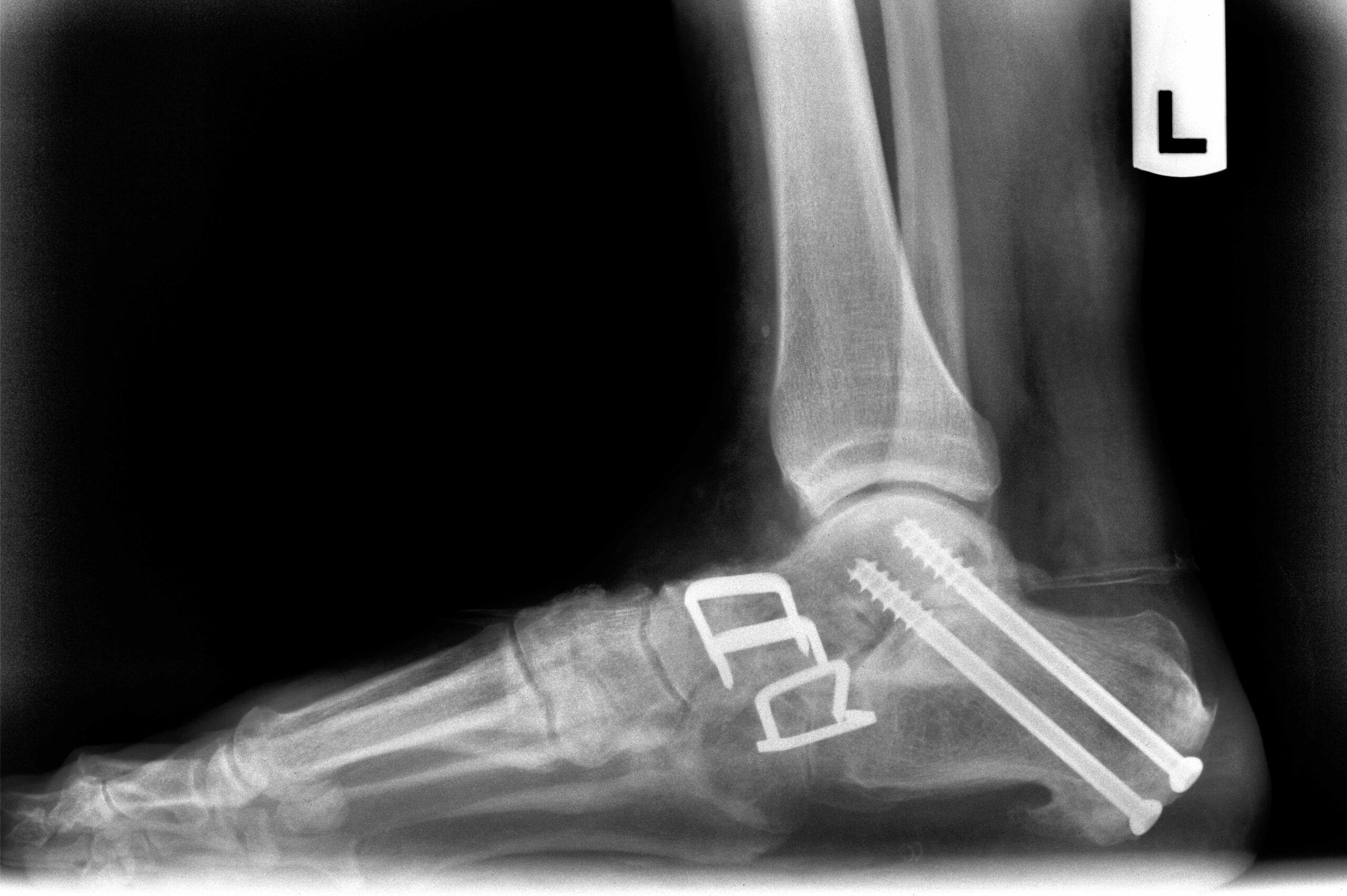
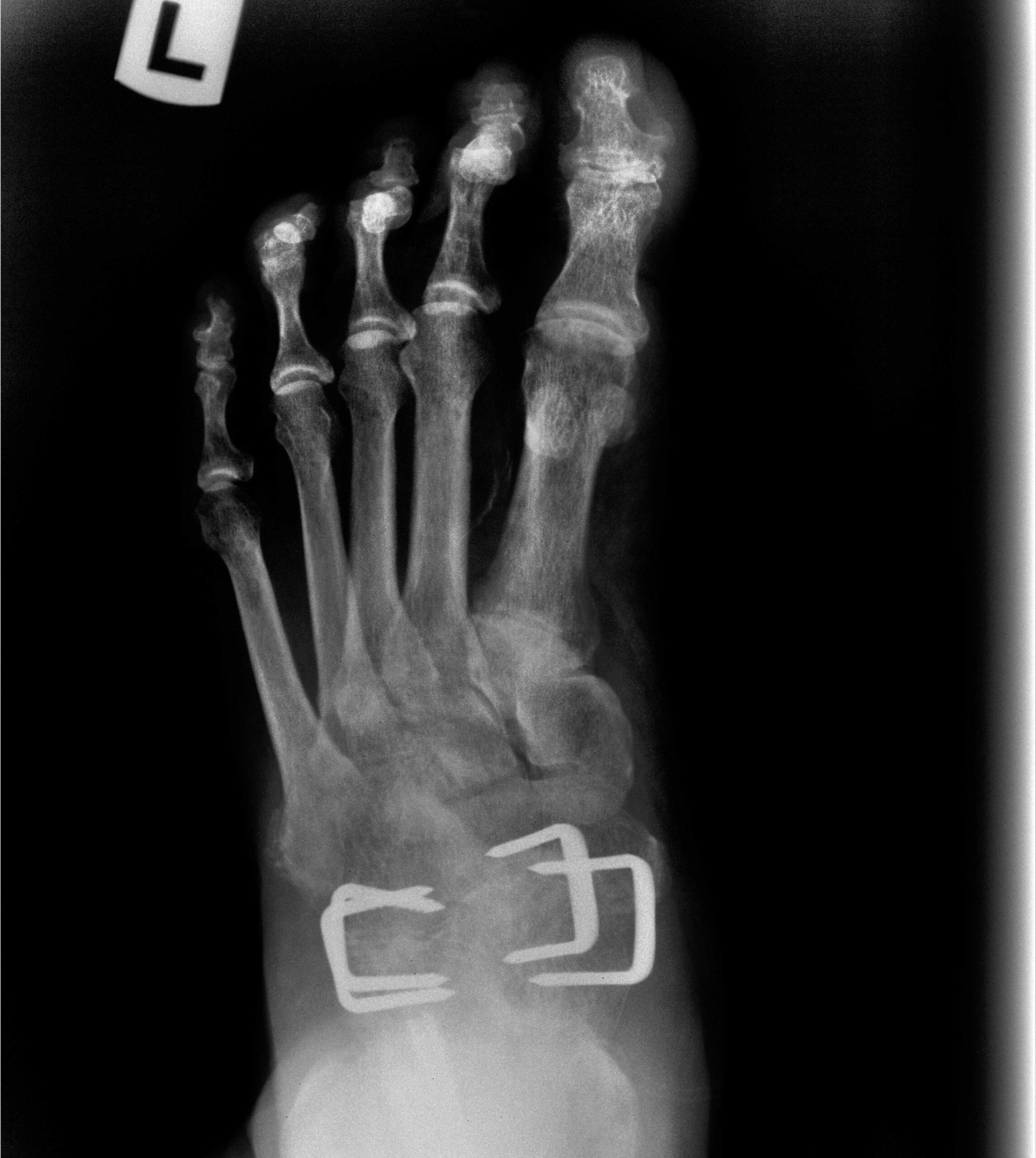
Post-operative films show restored talar declination and calcaneal pitch, screw fixation across the subtalar joint, and staple fixation reducing and holding the talonavicular joint. Individual anatomy, fixation choice, and healing course vary by patient — shared for educational purposes only.
Meet Dr. Kevin Lam, DPM, FACFAS, DABLES, DABPS
Training & Credentials
- Doctor of Podiatric Medicine with Honors — Temple University School of Podiatric Medicine
- Foot & Ankle Surgery training — Jackson Memorial Health System, Level 1 Trauma Center
- Podiatric Surgical Chief Resident — Mount Sinai Medical Center, Miami Beach
- Triple board-certified in Foot Surgery, Reconstructive Rearfoot & Ankle Surgery
- President, Family Foot & Leg Center, P.A. (since 2005)
Teaching & National Recognition
- Fellowship Director — FFLC Reconstructive Foot & Ankle Surgical Fellowship
- Adjunct Professor — Barry University and Temple University Schools of Podiatric Medicine
- National lecturer on reconstructive rearfoot and ankle surgical technique
- Named Among America’s Top Podiatrists / Top Doctors in Southwest Florida in multiple years since 2011
Dr. Lam’s reconstructive practice focuses on staged treatment of PTTD — from early tenosynovitis managed conservatively, through end-stage, fixed adult acquired flatfoot deformity requiring triple arthrodesis. That case mix, combined with a fellowship program he personally directs, is the kind of concentrated, teachable expertise in flatfoot reconstruction that is genuinely uncommon in Southwest Florida.
Already Been Told You Need a Flatfoot Reconstruction? Get a Second Opinion First.
Triple arthrodesis is a major, largely irreversible procedure. If you’ve been diagnosed with PTTD or adult acquired flatfoot and told you need surgery, get a second opinion from Family Foot & Leg Center before you sign a consent form. There is no cost to being certain.
Get Your Flatfoot Evaluated — Or Get a Second Opinion First
Nine Southwest Florida locations — Naples, Estero, Fort Myers, Cape Coral, Port Charlotte, and Sarasota. Same-day and next-day appointments are usually available.
This page is educational information, not a diagnosis. Individual treatment plans, staging, and surgical candidacy are determined in consultation with Dr. Lam. Weight-management therapies, including GLP-1 medications, should only be pursued under the guidance of a treating physician.